What is Intravascular Ultrasound tset (IVUS)?
Intravascular Ultrasound (IVUS) is a catheter-based diagnostic procedure used to view the inside of a coronary artery, providing a real-time view. IVUS shows the degree of narrowing or thickening (stenosis) of an artery by providing a visual image of the inside of the artery (the lumen) and the atheroma (membrane/cholesterol loaded white blood cells) that are hidden within the artery wall.
Dr. Augusto Villa use IVUS to image the lining of an artery in preparation for, during or to review the results of an angioplasty or arthrectomy. It is also used in the placement of stents.
Advantages of Intravascular Ultrasound Test
IVUS enables Dr. Villa to get inside the artery with a camera-like device. IVUS can quantify the percentage of narrowing and give insight into the nature of the plaque. It also may reveal what in the past has been referred to as “re-stenosis” (a recurrence of the plaque buildup that may have previously been removed). There is evidence that this is not a re-stenosis but rather the IVUS”s ability to see buildup that may have been missed during an angiogram and angioplasty.
What to Expect During Intravascular Ultrasound Test
IVUS is an invasive procedure and, as such, comes with the risks associated with any invasive procedure.
The entire procedure might take less than an hour or as long as several hours:
- An area around wrist (radius) or the groin will be shaved and cleaned in preparation for the insertion of a catheter (a thin tube).
- A mild sedative is administered to aid in relaxation.
- A local anesthetic is injected into the catheter site.
- Dr. Villa directs this catheter, painlessly, through arteries until it reaches the area to be studied.
- A guide wire with an ultrasound probe on its tip is inserted into the catheter and guided to the furthest position to be imaged.
- Sound waves are emitted from the probe. The probe also receives and returns the echo information, sending images to a computer.
- The guide wire is held in place and the probe is slid backwards – usually under steady, smooth, motorized control – sending and receiving ultrasound images along the way.
After the Intravascular Ultrasound Test
After the catheter is reomoved, patients must lie flat for two to six hours.
If a patient lives more than an hour”s drive from the hospital, they may need to stay overnight. Due to the administration of a mild sedative, patients will not be allowed to drive themselves home. If a patient arrives without a companion to take them home – and they are not staying overnight – the procedure will be cancelled and rescheduled.
Results of the Intravascular Ultrasound Test
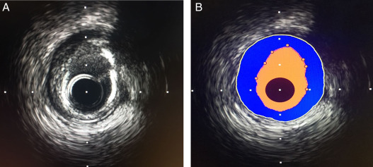
The blood vessel wall inner lining, atheromatous disease within the wall, and connective tissues are echogenic (they return echoes making them visible on the monitor). Blood and healthy muscular tissue are echolucent (they return no images, just black spaces on the monitor).
Heavy calcium deposits are very echogenic, which means they reflect sound, and are distinguishable by shadowing. Heavy calcifications are reflected as bright images with shadowing behind it.
Patients need to meet with Dr. Villa to discuss their test results and any recommended treatment plans.